Day 7: Goodb-EYE

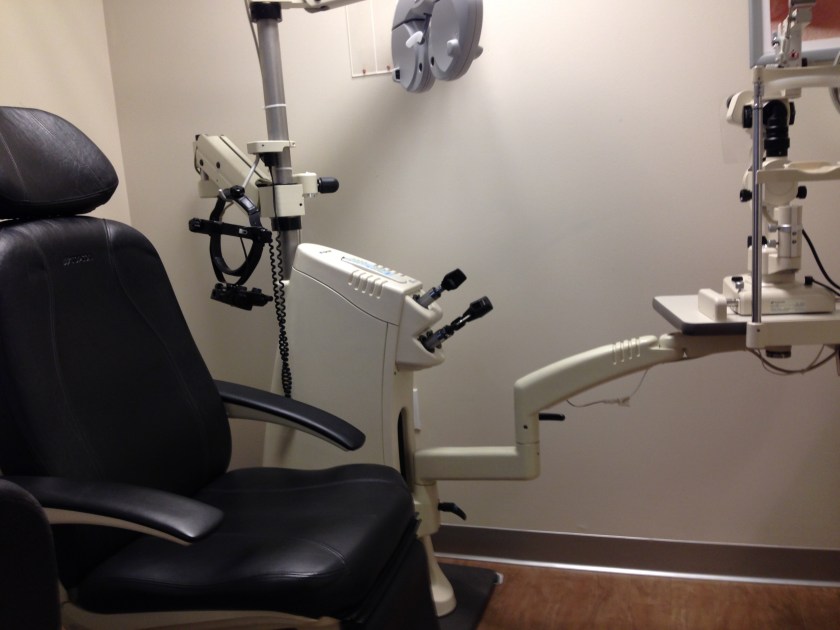
This was my final day at the Eye Center and even though I had to say goodbye, I didn’t leave without learning some new things. Through shadowing I got to see orbital fractures, hordoleum, and even ocular birthmarks. Hordoleum is an eye infection characterized by swelling on the eye lid or an isolated bump on the eye lid. There was also a case of ocular neoplasm which is pretty much a lesion on the retina. These are usually benign. I got to observe a technician under training to end the day and the experience. Since my experience has become whole, I decided to include a picture that shows the entire exam chair with all of the machinery.

image.jpeg


Day 6: Contacts

I returned to the Southpoint location today and continued to shadow Dr. Besharat. There were a lot of patients who came in for contact lenses today. Through shadowing I got learn about the distinct differences between using glasses and contact lenses. Glasses are usually more effective than contacts for two reasons. The first is that a contact lens makes contact with the surface of the eye which can lead to dryness and irritation on the eye. Dryness and irritation ultimately lead to a compromise of a patients vision. If not managed correctly dryness can lead to clouded vision and ultimately the contact lenses cannot be used again and the patient will have to go back to glasses. The second reason for the change in disparity is because of astigmatism. Astigmatism is irregularity in he curverture of the surface of the eye. Instead of being one smooth curve, the eye has varying areas of sloping. These variances lead to light entering the eye in irregular way and vision to be compromised. Glasses can be custom made to a persons eye so a correction for astigmatism can be added. The shape of a contact lens is standard and there are limitations to the amount if astigmatism correction that can be added to the prescription. In general a contact lens and glasses have different prescription because the lens sits on the surface of the eye whereas glasses sit on the ridge of the nose so there is a gap between the correction and the eyes. The below photos are of the machinery used to read the glasses,  evaluate and rectify the prescription, and also of a case with lenses in them that can be used to mimic the effect of a contact lens:

Day 5: Cary Towne Center

T0day I had the opportunity to work out of the Cary Towne Center location. I got to shadow Dr. Besharat again, but today I got more insight on the practice’s protocol for pediatrics. Many of the machines they use on a average age human being are either too complicated or just too big for a small kid to use. They have to use basic technology on children. This includes using a book to test for colorblindness and using pictures instead of letters to adjust the child’s prescription. Additionally, they use a slightly different solution for dilation so that it can absorb through the child’s eye lid and the child won’t feel anything on their eye. From an anatomical standpoint, the biggest difference between an adult eye and a children’s eye is that the child has an extra layer around their retina. This extra layer is almost like saran wrap in that it is clear and it is like a film that is wrapped around the retina. This usually peels off as the eye develops with age. I also got to watch diagnoses of angle close glaucoma, blepharitis, as well as stage 2 cataracts. Up to this point no patient has had any of these conditions. I also got to observe a patient that had degeneration of the cornea. In this condition, the cells of the cornea peel off due to extreme dryness of the eye. The usual treatment is to use lubricating drops as well as a bandage type contact lens temporarily to avoid pain. In the long term, a corneal specialist will need to be seen. The below pictures are of the sign on the outside of the Cary practice and of the less complex machinery Dr. Besharat uses in a pediatric setting to evaluate the inside of the eye and the overall health of the eye.

Day 4: The Prescription

I apologize for how late this post is, it is meant to be for Thursday May 26. Due to cross country travel and the busyness of a speech and debate tournament, I was slightly delayed posting.

This morning Ms. Allen payed us a visit at the Eye Center. We got to talk to her about the experience and finally gave her a tour of the center. After that I continued to shadow Dr. Besharat and I watched her conduct some tests she did conduct previously. She used a dye on the eyes of her patients to check for abrasions and other imperfections on the surface of the eye. Afterwards she conducted an exam on me and went through all of the standard procedures from checking the strength of my eye muscles to checking the health of my eye. Everything checked out! She also tested my prescription and I learned that I needed a new prescription which I was given. Finally, I was able to see Dr. Besharat check patients for potential tumors on their eyes. The below image is of the many bottles of dyes and solutions the doctor’s use on a daily basis. Some are lubricating drops, others are to control allergies, and others are for contact lenses. One of the bottles contains the dye in it.

IMG_1611

Day 3: My Own Pre-Screening

Today I had the opportunity to learn how to operate the pre-screening machinery in the office. I got try them on one of the technicians in the office. After that, the technician created a chart in the office’s system for me and she did a pre screen on me. From there I learned what all of the tests look for and a little bit about how to read the results. I also got retinal photographs taken. I did some more shadowing today and got to take a look at there contact lens protocol the office uses and their dedicated contact lense room where they keep their lens samples and where patients can try on different lenses. The below picture is of the room:

image

Day 1: The Pre-Screening

Unfortunately I did not see Dr. McElreath’s email until returning home, I was kept quite busy today! I shadowed the optometric technicians today and watched them go through their normal test protocols. They conduct vision tests both with and without corrections (contact lenses or glasses). They conduct color vision tests without the correction and the depth perception test with correction. They also, if the patient agrees to doing it, take retinal photographs which can allow the optometrist to diagnose the patient with diseases such as glaucoma and eye infections. These photos can also help diagnose diabetes and hypertension. I also watched the technicians go through their process of interviewing the patients. They ask about a variety of things ranging from family medical history to how old a prescription is. I also got to watch how the receptionists in the front checked patients in and out and how information about a given patient is communicated from a technician to the doctor and vice versa. — Viraj

IMG_1597.JPG

Day 2: The Exam

Today was another busy day at the Eye Center at Southpoint Mall. I got to shadow Dr. Besharat today and got to see how she conducts her eye exams. The majority of the exams begin with checking the patients range of motion and then checking their prescription. Finally, Dr. Besharat asks the patient about any specific concerns they may have. Dr. Besharat diagnosed patients with glaucoma, cataracts, central serous retinopathy, and choroidal nevus. Glaucoma and cataracts are very common in patients and the other two conditions not so much. Central serous retinopathy is the clouding of macula that leads to blurred vision in usually one eye, it is caused by stress. A choroidal nevus is nothing more than a freckle on the eye that can sometimes cause a noticeable floater and other times can go unnoticed. It is very interesting how many unnoticed problems people have in their eyes and they think everything is OK because their prescription is working fine and they are able to see. I also got to see routine exams for updating prescriptions and changing contact lenses. At the end of the day I got to follow the entire appointment process from pre-screening to actually seeing the doctor. I also got to learn about their flagging system for their exam rooms. Below are photos of an exam chair, the actual machine used during an exam, and the flags near an exam room. — Viraj

IMG_1602IMG_1604.JPGIMG_1605.JPG